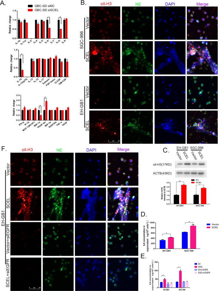
Fig. 4. SCEL influence the neutrophil extracellular traps formation.
A The indicated cells conditioned medium were performed liquid suspension array. B Neutrophils were isolated from peripheral blood coculture with the indicated GBC cells for 3.5 h and then representative immunofluorescent staining photographs of cit-histone3 (red), NE (green) and DAPI (blue) are shown by fluorescence microscope. C Cit-histone H3 protein levels were determined by Western blot, grayscale statistics at the bottom of Western blot band. D ELISA were performed to verify the regulatory effects of SCEL on IL8 in indicated cell lines. E, F The indicated GBC cell lines were incubated with or without EGFR siRNA and then subjected to ELISA array to detect the IL8 concentration (E) or cocultured with neutrophil to identity the neutrophil extracellular traps formation (F). *p < 0.05, **p < 0.001, ***p < 0.001.

