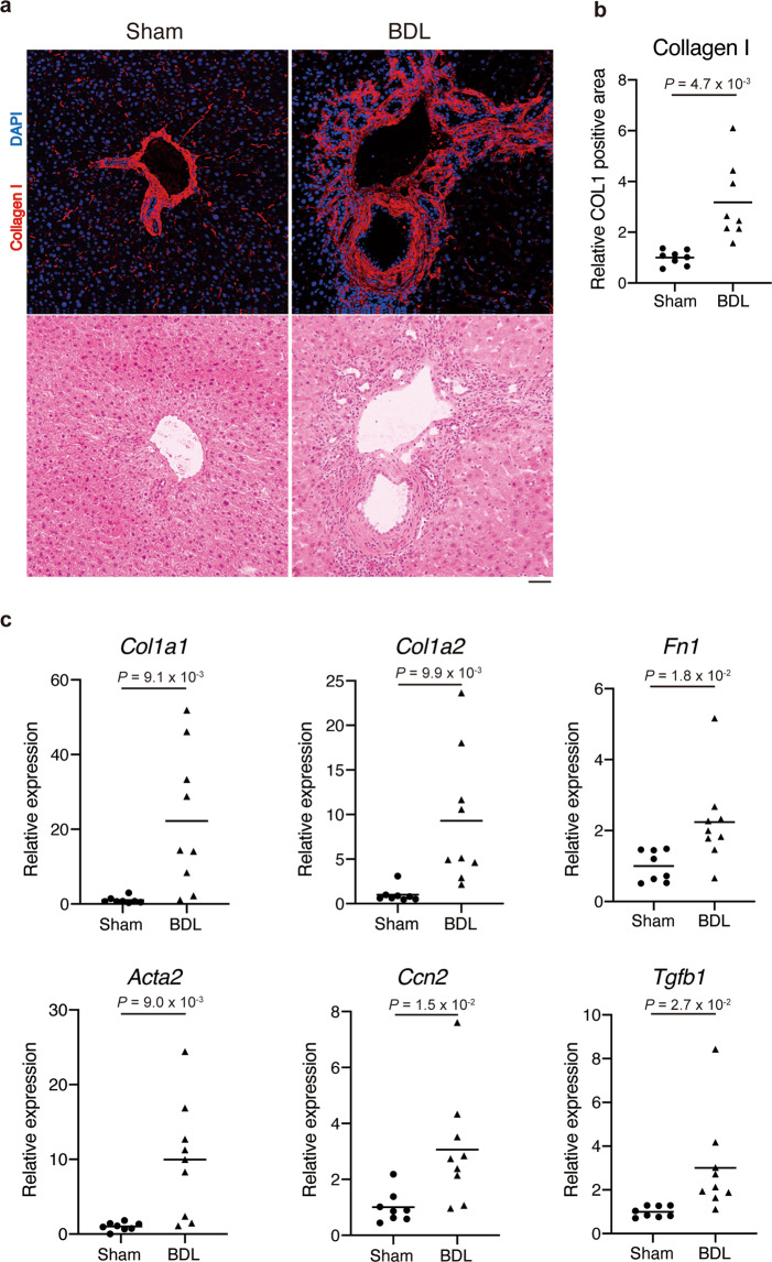
Fig. 1. BDL induces liver fibrosis in mice.
The common bile duct of eight-week-old male mice was ligated to induce liver fibrosis. a The sections derived from livers excised on day 7 post-operation were subjected to immunofluorescence staining for collagen I and DAPI (upper) and subsequently to H&E staining (lower). b The collagen I-positive area was quantified. n = 8 mice for each group. c The hepatic expression levels of fibrosis-related genes (Col1a1, Col1a2, Fn1, Acta2, Ccn2, and Tgfb1) were quantified on day 7 post-operation. n = 8 mice for sham and n = 9 mice for BDL. Data represent individual data points and the means. Data were analysed using the two-sided unpaired t-test and Welch’s correction. Scale bar, 50 μm (a).

