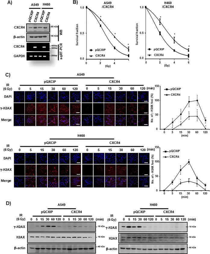
Fig. 3. CXCR4 upregulation enhances IR resistance in NSCLC cells.
A Western blot (WB, upper) and RT-PCR (lower) analysis of NSCLC cells (A549 and H460) transfected with CXCR4. β-actin and GAPDH were used as a loading control for Western blot and RT-PCR, respectively. B IR clonogenic survival assay of control vector (pQCXIP) and CXCR4 overexpressing (CXCR4) NSCLC cells. Cells were exposed to IR at the indicated dose in the Figure. C γ-H2AX foci assay of A549 (upper) and H460 (lower) cells. Cells were transfected either with control vector (pQCXIP) or pQCXIP-CXCR4 (CXCR4) and were exposed to IR (6 Gy) and fixed at the indicated time points in the Figure. Representative ICC images of γ-H2AX (left) and quantification of the results (right). Nuclei were counterstained with DAPI (blue). White bar: 20 μm. *P < 0.05, **P < 0.01. D Western blot analysis of A549 (left) and H460 (right) cells exposed to IR (6 Gy) at the indicated time points transfected with control vector (pQCXIP) or pQCXIP-CXCR4 (CXCR4). H2AX and β-actin were used as loading controls.

