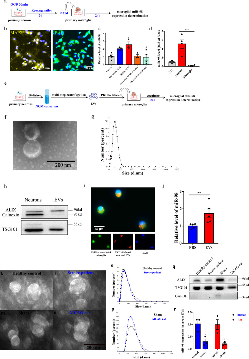
Fig. 2. MiR-98 packed into EVs can be transferred from neurons to microglia.
a The procedure of primary neuronal-conditional media (NCM) collection and coculture with primary microglia. b Positive rate of Rat primary cortical neuron (MAP2) and microglia (IBA1). c The relative miR-98 expression level of microglia after added with NCM from starvation and OGD-R treatment and GW4869 blocking. (n = 3 independent experiments). d The relative miR-98 expression level between neuron and microglia of purification culture, using the miR-98 expression of Neuro-2a (N2a) cells as a baseline. e The procedure of extraction of primary neuronal EVs and co-cultured with microglia. f Transmission electron micrograph (TEM) of neuronal EVs. g Size distribution of neuronal EVs based on Zetasizer Nano-ZS90 measurements. h Western blot for EVs markers Alix, TSG101, and Calnexin. i Representative images of internalization of PKH26-labeled neuronal EVs into the CellTrackerTM Green-labeled microglia 12 h after coculture. j Relative miR-98 level in microglia 24 h after incubated with PBS and neuronal EVs (extraction by ~80~100 ml neuronal media). (n = 5 independent experiments). Data are shown as means±S.E.M., *P, #P < 0.05; **P < 0.01; ***P < 0.001 vs corresponding control. k–n Transmission electron micrograph (TEM) of serum EVs from both individuals and rats. o–p Size distribution of serum EVs from both individuals and rats. q Western blot for EVs markers Alix, TSG101, and GAPDH. r The relative miR-98 expression level between in serum EVs between both individuals and rats.

