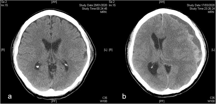
Fig. 1.
a Computed tomographic (CT) scan of the brain of a 59-year-old gentleman at the time of injury. He had a minor head injury with retrograde amnesia. CT scan showed a left-sided scalp hematoma but no intracranial hemorrhage and he was discharged with medical advice. Subsequently, he had a progressively worsening headache. b He finally re-attended the hospital 7 weeks after the initial minor head injury. The new CT showed a left-sided chronic subdural hematoma with mass effect and midline shift. He had a good recovery after emergency burr hole drainage

