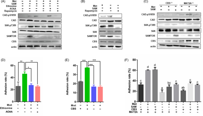
FIGURE 8.

Methionine promoted embryo proliferation and implantation. A, JAR cells were starved overnight in serum and methionine‐free medium. Cells were treated with methionine, SAM, ethionine, AOAA and rapamycin, and then collected for Western blot analysis. B, JAR cells were pre‐treated with rapamycin for 12 h in order to inhibit mTORC1. Afterwards, the culture medium was discarded and cells were rescued with methionine and SAM. Cells were collected for Western blot. C, CBS or MAT2A was silenced in JAR cells and then treated with methionine and SAM. Cells were collected for Western blot analysis. D, JAR and Ishikawa cells were starved overnight in serum and methionine‐free medium and then treated with methionine, SAM, ethionine, AOAA, (E) CBS silence and (F) methionine, SAM, MAT2A/CBS silenced. Then, JAR cells were stained with CFSE and plated on an Ishikawa monolayer cells. Implantation rates were measured using flow cytometry. n = 3. ***P < .001; **P < .01; *P < .05
