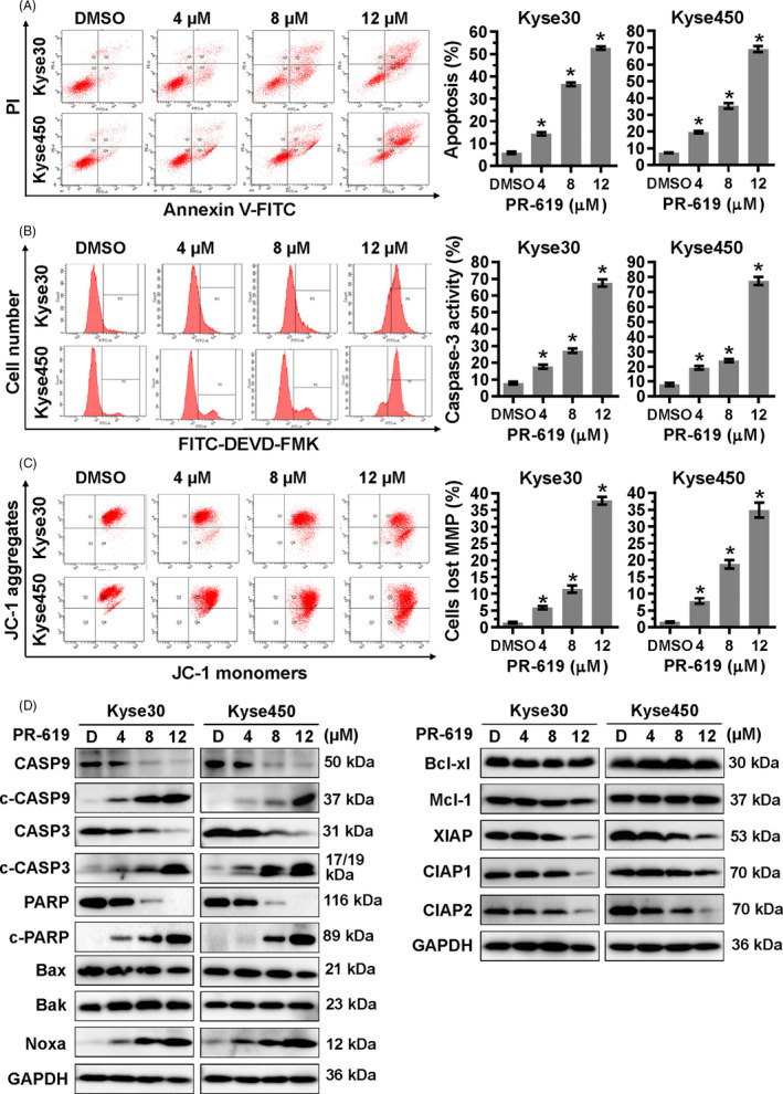
FIGURE 2.

PR‐619 induced intrinsic apoptosis. Kyse30 and Kyse450 cells were treated with PR‐619 for 48 h. A, Apoptosis was determined by FACS analysis using Annexin V‐FITC/PI double‐staining kit (left panel), and Annexin V+ cell populations were defined as apoptosis (right panel). B, CASP3 activity was also analysed with FACS (left panel). The percentage of cells with active caspase3 was shown in the right panel. C, PR‐619 induced mitochondrial membrane depolarization. Cells were treated with PR‐619 and analysed by FACS as described in Materials and Methods section. D, Effect of PR‐619 on the expression of apoptotic proteins, pro‐apoptotic and anti‐apoptotic proteins. Kyse30 and Kyse450 cells were treated with PR‐619 for 48 h, and cell extracts were prepared for Western blotting analysis. GAPDH was used as the loading control. All data were representative of at least three independent experiments (n = 3; error bar, SD)
