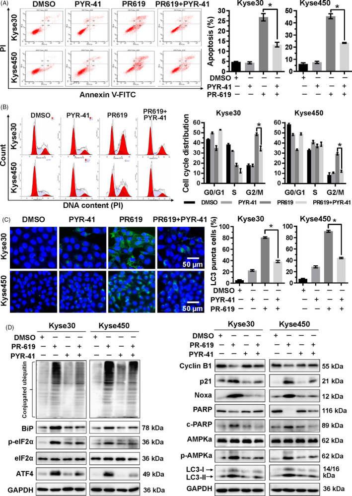
FIGURE 7.

Accumulation of ubiquitinated proteins participated in the cell growth inhibition effect of PR‐619. A, PYR‐41, ubiquitin E1 inhibitor, increased PR‐619 induced apoptosis. Kyse30 and Kyse450 cells were treated with PR‐619 (8 µmol/L) single or combined with PYR‐41 (20 µmol/L). Apoptosis was determined by FACS analysis using Annexin V‐FITC/PI double‐staining kit (left panel) and Annexin V+ cell populations were defined as apoptosis (right panel). B, PYR‐41 decreased G2/M cell cycle arrest. Kyse30 and Kyse450 cells were treated with PR‐619 single (8 µmol/L) or combined with PYR‐41 (20 µmol/L), followed by PI staining and FACS analysis for cell cycle profile (left panel). Distribution was analysed by Modifit and GraphPad software (right panel). C, PYR‐41 reduced PR‐619 induced autophagy. Kyse30 and Kyse450 cells were treated with PR‐619 single (8 µmol/L) or combined with PYR‐41 (20 µmol/L). LC3 was detected using immunofluorescence assay, reprehensive pictures were captured (left panel), and LC3 puncta cells were statistically analysed (right panel). D, Effect of PYR‐41 on protein expression of ER stress, cell cycle‐related proteins, Noxa, c‐PARP, PARP, p‐AMPKα, AMPKα and LC3. Kyse30 and Kyse450 cells were treated with PR‐619 single (8 µmol/L) or combined with PYR‐41 (20 µmol/L). Cell proteins were detected using specific antibodies. GAPDH was used as the loading control. All data were representative of at least three independent experiments (n = 3; error bar, SD)
