Figure 3.
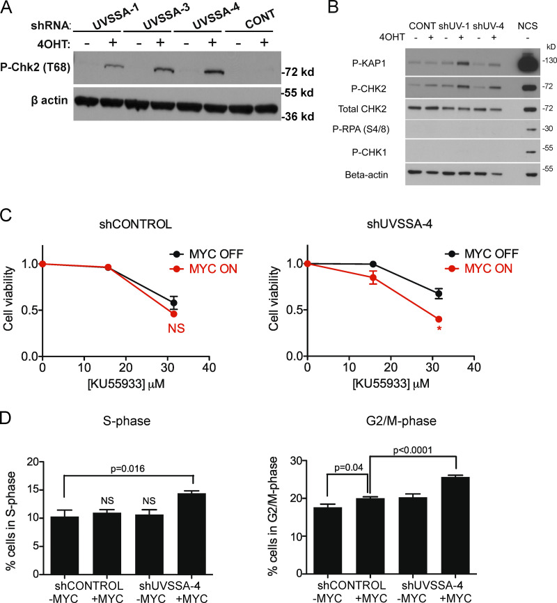
Down-regulation of UVSSA elicits a DNA damage response in MYC-deregulated cells. (A) CHK2 activation in three independent shUVSSA cell lines following MYC induction. Indicated cell lines were treated with 200 nM 4OHT or vehicle and 1 µg/ml doxycycline every 3 d and harvested at day 9. Whole-cell lysates were subjected to Western blot analysis with anti–phospho-T68-CHK2 antibodies. β-Actin staining served as a loading control. (B) KAP1 phosphorylation in two independent shUVSSA cell lines following MYC induction. Indicated cell lines were treated with 200 nM 4OHT or vehicle and 1 µg/ml doxycycline every 3 d and harvested at day 9. Whole-cell lysates were subjected to Western blot analysis with indicated antibodies. β-Actin staining served as a loading control. NCS treatment served as a positive control for induced DNA DSBs. (C) MTS assays in the presence of ATM inhibitor KU55933. shCONTROL (left) or shUVSSA-4 (right) cells were subjected to standard MTS assays with increasing concentrations of KU55933. Asterisks indicate significant difference from MYC off at indicated concentration (*, P < 0.05). Error bars represent the SEM (n = 3). (D) Cell cycle distribution of shCONTROL or shUVSSA-4 cells in MYC on or MYC off conditions. Cells in S phase (left) or G2/M phase (right) were measured by propidium iodide staining after 30 d of culturing cells with 1 µg/ml doxycycline and 200 nM 4OHT or vehicle. P values are indicated where appropriate. Error bars represent the SEM (n = 3).

