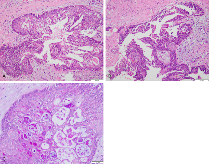
Figure 2.

Histopathologic features of mucoepidermoid carcinoma of the breast. A. The lesions are constituted by solid neoplastic nests and scattered cystic spaces filled with mucoid material; (×100). B. Three cell types were observed in mucoepidermoid carcinoma. Intermediate cells are on the left (red arrow); epidermoid cells are localized centrally (black arrow); (×100). C. AB-PAS stains show numerous mucinous cells in the invasive component (The arrow points to mucoid material); (×100).
