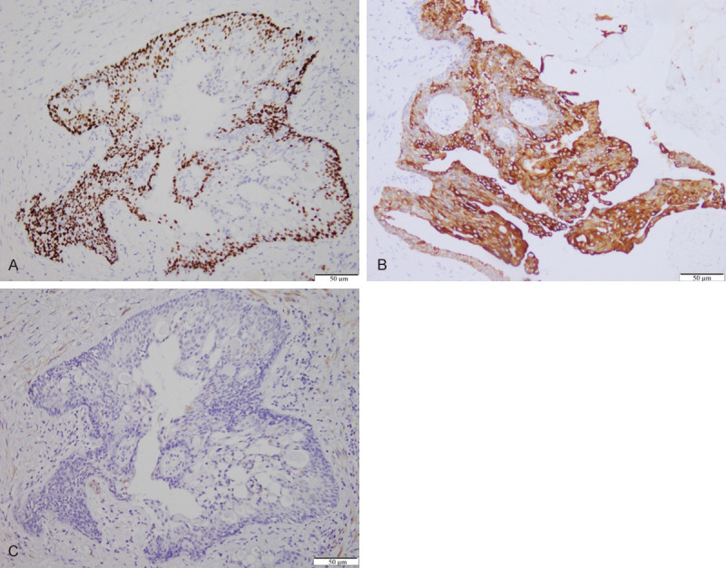
Figure 3.

Immunohistochemistry of mucoepidermoid breast carcinoma. A. Epidermoid and intermediate cells are positive for p63, whereas mucinous cells around the microcysts and toward the cystic lumen are mostly negative (The arrow points to a positive stain); (×100). B. Glandular cells are positive for CK 7 (The arrow points to a positive stain); (×200). C. Tumor cells are negative for calponin; (×100).
