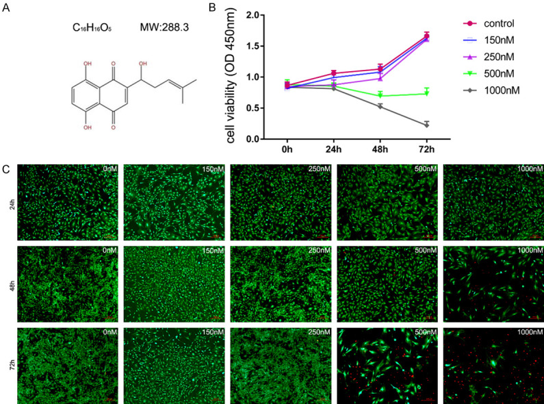
Figure 1.

(A) The molecular structure of shikonin. High concentration of shikonin restrained the proliferation of BMSCs. Primary BMSCs were treated with shikonin before measuring cell viability. The CCK-8 assay (B) and live/dead staining (C) showed that the number of dead BMSCs decreased significantly after 48 hours of shikonin treatment at a concentration higher than 250 nM. Cytotoxicity (live/dead) assay images show the live (green) and dead cells (red) in a cellulose sample.
