Figure 1.

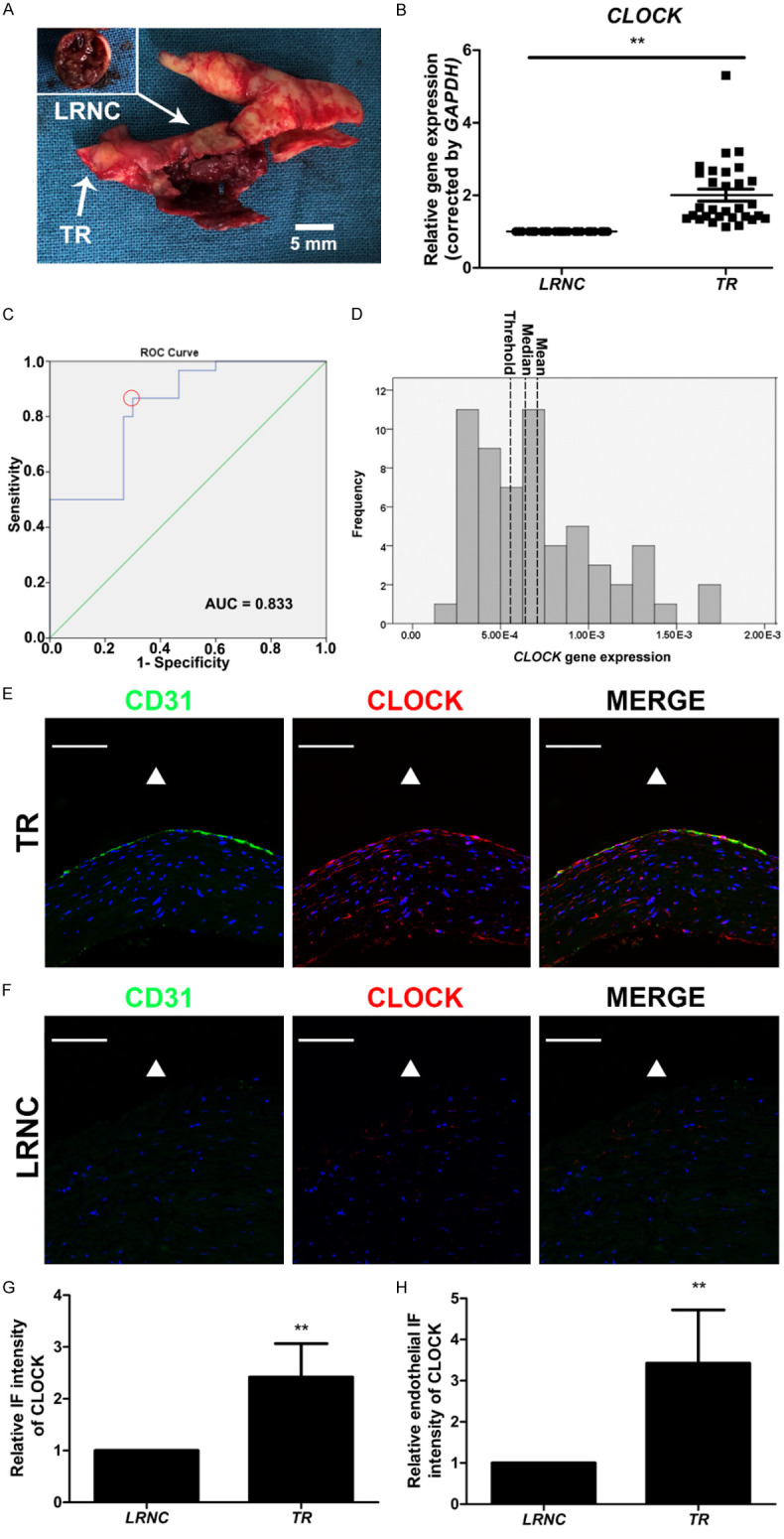
Decreased CLOCK mRNA levels were associated with the development of carotid atherosclerotic plaques. A. Morphology of the lipid-rich necrotic core (LRNC) with intraplaque hemorrhage and transitional region (TR) of carotid atherosclerotic plaque. B. CLOCK mRNA levels were detected in LRNC (n = 30) and TR (n = 30) of tissues from patients with internal carotid artery stenosis. C. Receiver-operating characteristic curve of CLOCK mRNA levels and TR (red dotted circle, optimal cut-off point). D. Histogram of CLOCK mRNA levels (left dotted line, threshold, 5.56 × 10⁻⁴; middle dotted line, median, 6.38 × 10⁻⁴; right dotted line, mean, 7.09 × 10⁻⁴). AUC, area under curve. E, F. Immunofluorescence staining for CLOCK (red) and CD31 (green) of TR and LRNC. Nuclei were stained with 4’,6-diamidino-2-phenylindole (DAPI; blue). The triangle indicates the lumen side. G. Immunofluorescence intensity of CLOCK in the TR and LRNC (n = 5). H. Endothelial immunofluorescence intensity of CLOCK in the TR and LRNC (n = 5). Scale bar: 100 μm. **P < 0.01.
