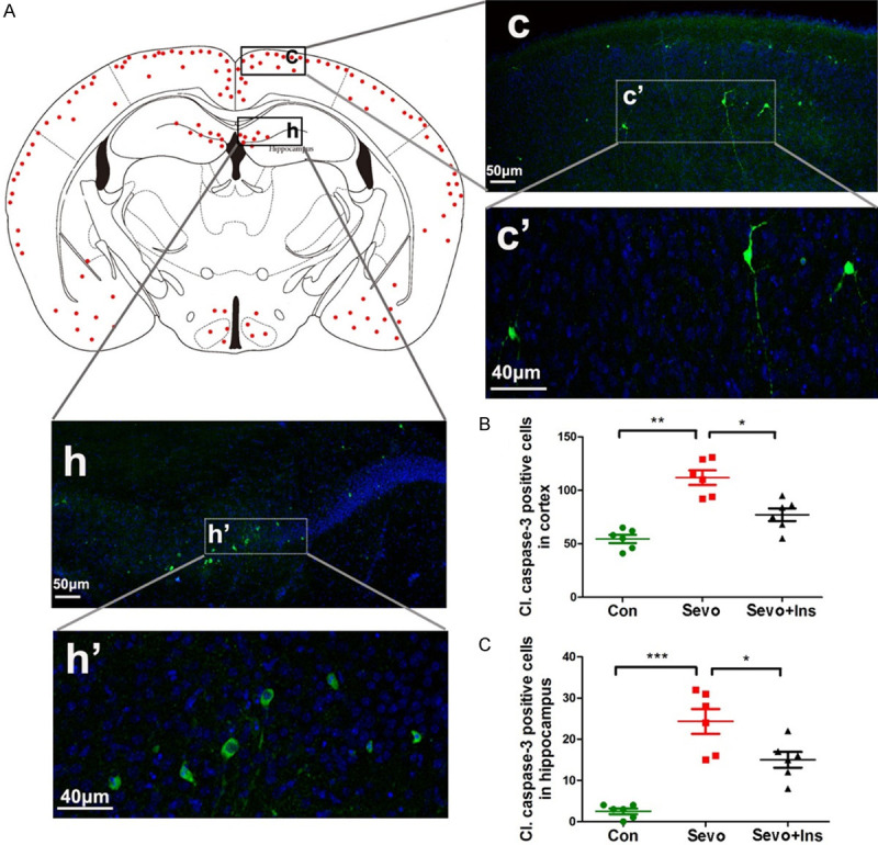
Figure 3.

Immunofluorescence of cleaved caspase-3 of neonatal mouse brains after sevoflurane exposure and intranasal insulin treatment. (A) P7 mice received intranasal administration of insulin or, as a control, saline, followed by inhalational anesthesia with 2.5% sevoflurane for 6 hr, beginning 30 min after intranasal administration. The mice were sacrificed at 6 hr post anesthesia, and the brains were fixed and immunostained by using antibody against cleaved caspase-3 (green) and counter-stained with nuclear marker TO-PRO (blue). The red dots in the mouse brain diagram in (A) represent the relative intensities of cleaved caspase-3-positive cells in various regions of the mouse brains after exposure to sevoflurane. (B, C) Neurons positive to cleaved caspase-3 in the cerebral cortex (B) and in the hippocampus (C) were counted separately and are shown as cleaved caspase-3-positive neurons per section (mean ± SEM, n = 6 mice/group). *, P < 0.05; **, P < 0.01; ***, P < 0.001, as analyzed using one-way ANOVA plus post hoc tests.
