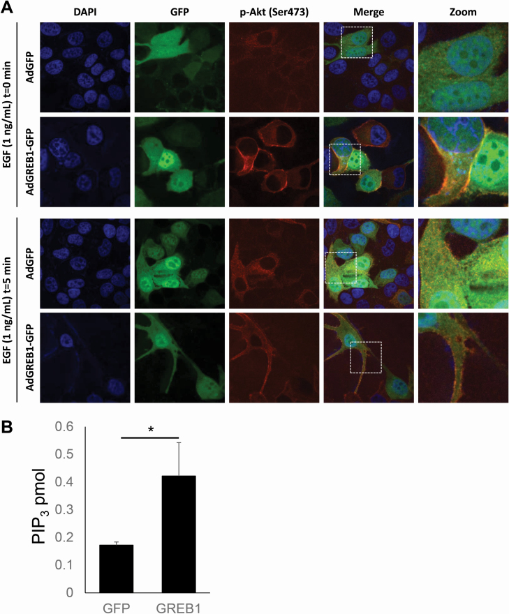
Figure 5.
Exogenous GREB1 promotes recruitment of Akt to the plasma membrane. (A) MCF7 cells were transduced with adenovirus expressing GFP or GREB1. The cells were then cultured in serum-free media for 16 h before being stimulated with 1 ng/ml EGF for 0 or 5 min. Cells were fixed and stained for 4′,6-diamidino-2-phenylindole or p-Akt (Ser473). Immunofluorescence microscopy was used to visualize the activation and localization of Akt. (B) MCF7 cells were transduced with adenovirus to express exogenous GFP or GREB1 and serum starved for 16 h. Lipids were extracted from all samples and levels of PIP3 were measured via enzyme-linked immunosorbent assay. Graphs represent mean PIP3 (pmol) + SD (n = 3). *P ≤ 0.05.

