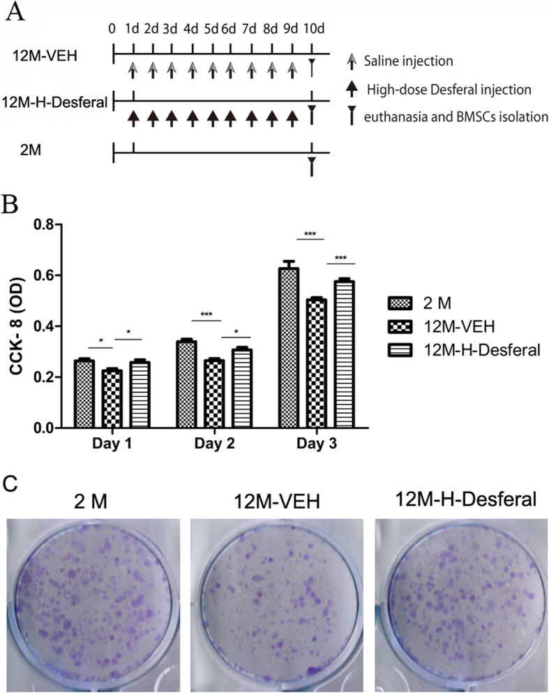
Fig. 3.

Cell proliferation and colony-forming of BMSCs from 2M, 12M-VEH and 12M-H-Desferal group rats. a Schema of short-term Desferal® administration of the ex vivo study. b CCK-8 assay of cell proliferation. The optical density (OD) value was measured at 450 nm absorbance. In BMSCs from 12M-H-Desferal® group, the proliferation rate (OD value) significantly increased compared with those from 12M-VEH group. While the OD values of 12M-VEH group are lower than those from 2M group at day 1–3. The data were drawn from three independent experiments and the results were expressed as mean ± SD. *p < 0.05, ***p < 0.001. c Colony-forming cell assay. Decreased cell colonies in BMSCs from the 12M-VEH group compared with that from the 2M group. While cell colonies in BMSCs from the 12M-H-Desferal® group increased significantly compared with that from the 12M-VEH group
