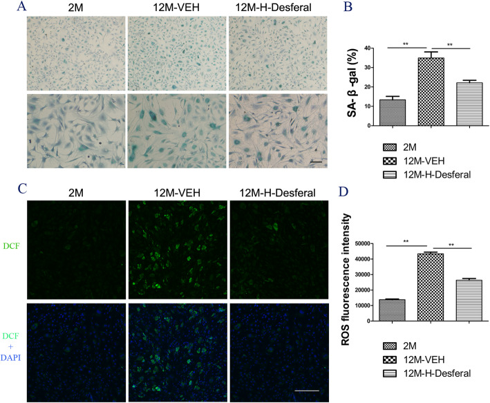
Fig. 5.
Changes of cell senescence and ROS in BMSCs from rats with and without Desferal® treatment. a Representative images of SA-β-gal staining. Blue stained cells indicates positive for senescence. Bar = 200 μm. b Quantitative assessment the percent of SA-β-gal positive cells. The percentage of senescence cell was significantly reduced in BMSCs from the 12M-Desferal® group compared with that from the 12M-VEH group. **p < 0.01. c Reactive oxygen species (ROS)-induced fluorescence in BMSCs at cell slides was visualized by confocal microscopy. ROS could react with a fluorescence sensor to produce a fluorescent product named as 2′, 7′-dichlorodihydrofluorescein (DCF), which is proportional to the content of ROS. Bar = 200 μm. d ROS quantification of BMSC on a 96-well plate by microplate assays. Decreased intracellular ROS levels were observed in BMSCs from the 12M-H-Desferal® group compared with that from the 12M-VEH group. **p < 0.01

