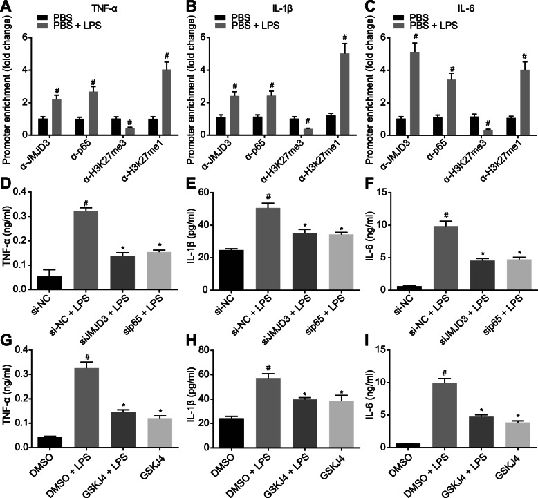
Fig. 3.
JMJD3 interacting with NF-κB/p65 elevates expression of the pro-inflammatory cytokines in LPS-induced BMDMs. a–c The recruitment of JMJD3, p65, H3K27me3, and H3K27me1 in the promoter region of pro-inflammatory cytokines TNF-α (a), IL-1β (b), and IL-6 (c) in BMDMs analyzed by CHIP-qPCR. #p < 0.05 vs. PBS-treated BMDMs. d–f The expression of LPS-induced pro-inflammatory factors TNF-α (d), IL-1β (e), and IL-6 (f) after knockdown of JMJD3 and p65. *p < 0.05 vs. PBS-treated BMDMs in response to si-NC; #p < 0.05 vs. BMDMs in response to si-NC. g–i The expression of pro-inflammatory factors TNF-α (g), IL-1β (h), and IL-6 (i) in BMDMs after 1-h treatment with GSK-J4 (4 umol/L, pharmacological inhibitor of JMJD3). *p < 0.05 vs. LPS-treated BMDMs in response to GSK-J4; #p < 0.05 vs. BMDMs in response to GSK-J4. The experiment was repeated 3 times independently. Quantitative data were presented as mean ± standard deviation. Unpaired data in compliance with normal distribution and equal variance between two groups were compared using unpaired t test. Comparisons among multiple groups were analyzed by one-way ANOVA with Tukey’s post hoc test. p < 0.05 indicated significant difference

