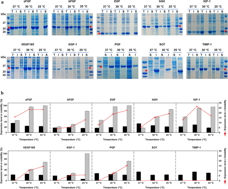
Fig. 2.
Expression of 6HFh8-GFs in flask culture. The proteins were induced by lactose in auto-induction media. They were then purified by HisTrap chromatography. a SDS-PAGE analysis of protein expression. Red dot, the fusion protein. I: insoluble fraction; S: soluble faction; T: total fraction. The images are representative of two independent experiments. b Quantitative analysis of protein expression. The bar graphs show percent expression (black bar) and solubility (gray bar) (primary y-axis), and the red line indicates expression level times solubility (secondary y-axis). Red line peak indicates the optimal temperature for fusion protein expression. Except for SCF and TIMP-1, all proteins were successfully expressed in soluble form at low temperature (25 °C and/or 30 °C). The expression level and solubility were analyzed by densitometry using ImageJ with two independent experiments

