Fig. 5.
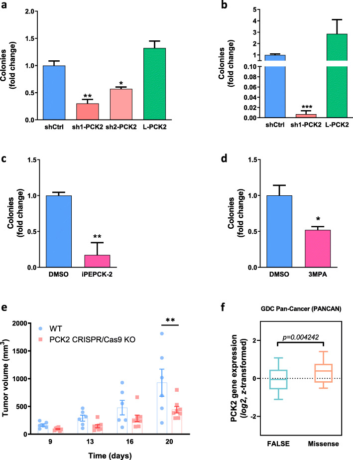
Effects of PEPCK-M activity change on anchorage-independent and xenograft growth. a–d Quantification of anchorage independent growth of HeLa and NIH-3T3Kras cells. Cells were seeded in semisolid agarose prepared with DMEM media 1:1 and grown for 4 weeks. At the end of incubation, cells were stained with MTT and number of spheres was counted. HeLa cells were grown in the presence of high-glucose media (25 mM) (a, c) or glucose exhaustion media (1 mM) (b). During the whole experiment, HeLa shCtrl cells in c were treated with PEPCK-M inhibitor (iPEPCK-2; 5 μM) or vehicle (DMSO). NIH-3T3Kras cells in d were grown in high-glucose media and treated with PEPCK-M inhibitor (3MPA; 100 μM) or vehicle (DMSO). Treatment media was refreshed every 3–4 days. Results are presented as fold change to shCtrl or DMSO, respectively. Statistical significance to shCtrl or DMSO was determined by unpaired two-tailed Student’s t test. e Tumor growth of SW480 WT and PCK2 CRISPR/Cas9 KO cells implanted into the flanks of BALB/C nude mice. Significance was determined using two-way ANOVA with Sidak multiple comparison post-test analysis. f Gene expression comparison analysis of tumor samples in the GDC Pan-Can dataset. Tumors presenting missense (Missense; n = 48) variant mutations are compared to non-variant tumors (FALSE; n = 11542). Unpaired t test analysis with Welch’s correction was used to determine statistical significance

