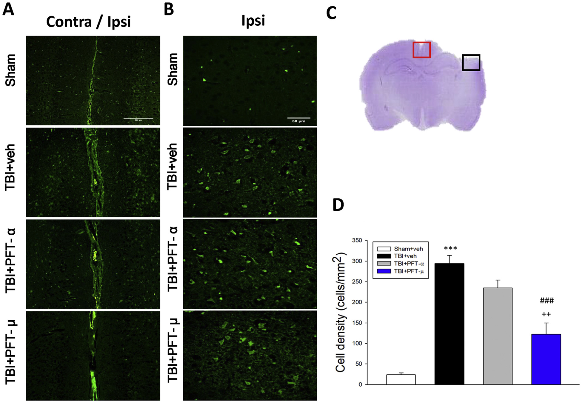
Fig. 3.

Post-injury administration of PFT-μ at 5 h after TBI significantly decreased FJC-positive cells at 24 h. FJC-stained (A) contralateral and ipsilateral (Bar = 200 μm) (B) ipsilateral cortex (Bar = 50 μm) regions of interest in sham, TBI + veh, TBI + PFT-α or TBI + PFT-μ group. (C) A representative HE-stained coronal section showing the area as indicated by the red (for both contralateral and ipsilateral cortex) and black (only ipsilateral cortex) square box to compare the fluorescent signals across the 4 groups of rats. (D) There was a significant decrease in the number of FJC-positive cells in the TBI + PFT-μ group. Data are expressed as means ± SEM. The total number of FJC-positive cells was expressed as the mean number per field of view (n = 5 per group). ***P < .001 versus sham group; ###P < .001 versus TBI + vehicle (veh) group; ++P < .01 versus TBI + PFT- α group.
