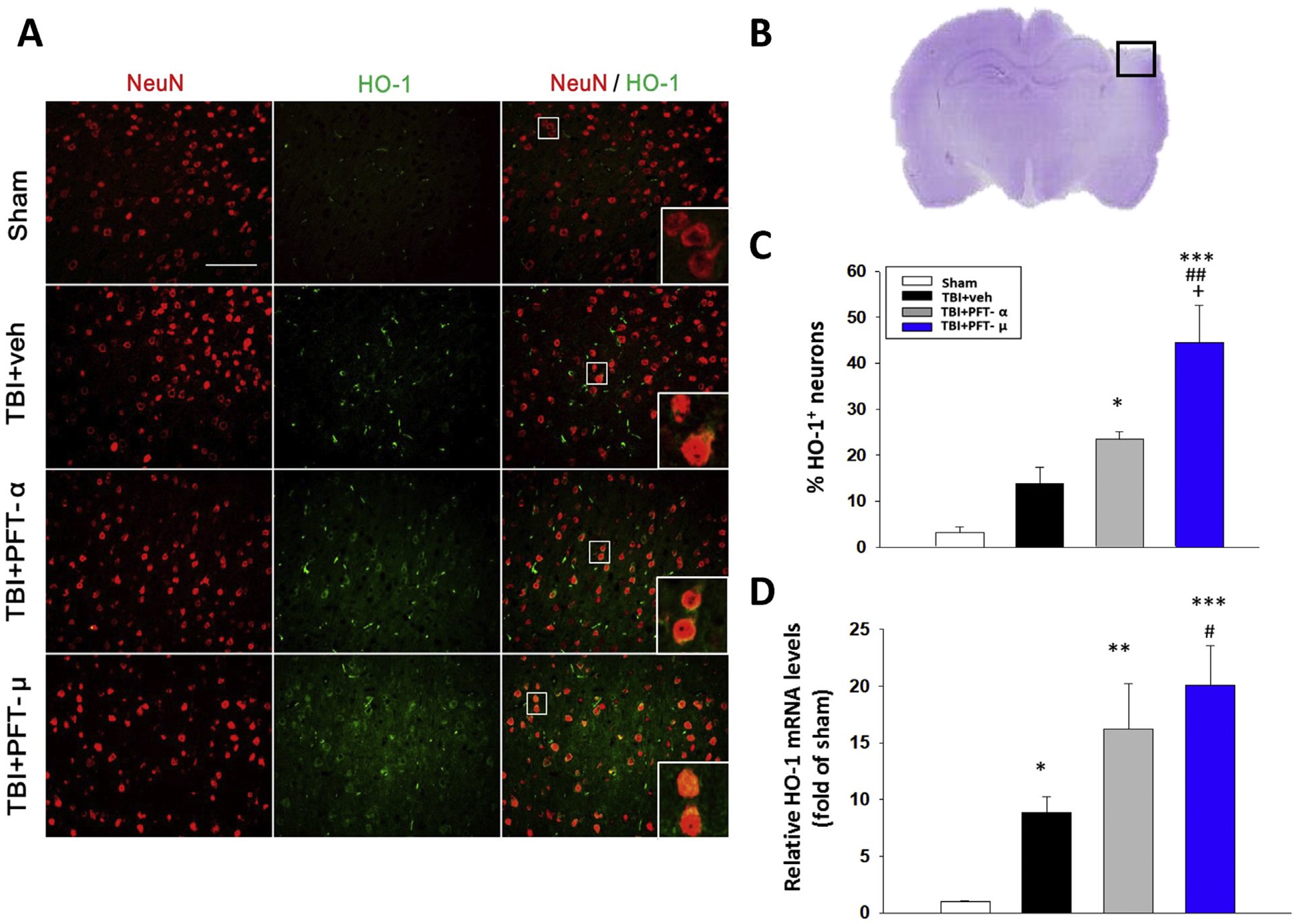
Fig. 5.

Post-injury administration of PFT-α and, in particular, PFT-μ at 5 h after TBI elevated HO-1 positive neurons and mRNA expression in the cortical contusion region at 24 h. (A) Co-immunohistochemistry of HO-1 and NeuN in cortical brain tissue. (B) A representative HE-stained coronal section showing the area as indicated by the black (ipsilateral cortex) square box to compare the fluorescent signals across the 4 groups of rats. (C) There was a significant increase of HO-1 positive neurons in the TBI + PFT-μ group beyond that induced in the TBI + vehicle group. (D) The mRNA levels of HO-1 were analyzed by RT-PCR. Data are expressed as means ± SEM (n = 5 per group). *P < .05, **P < .01, ***P < .001, compared with the sham group; #P < .05, ##P < .01, compared with the TBI + veh group; +P < .05, compared with the TBI + PFT-α group. Scale bar = 100 μm.
