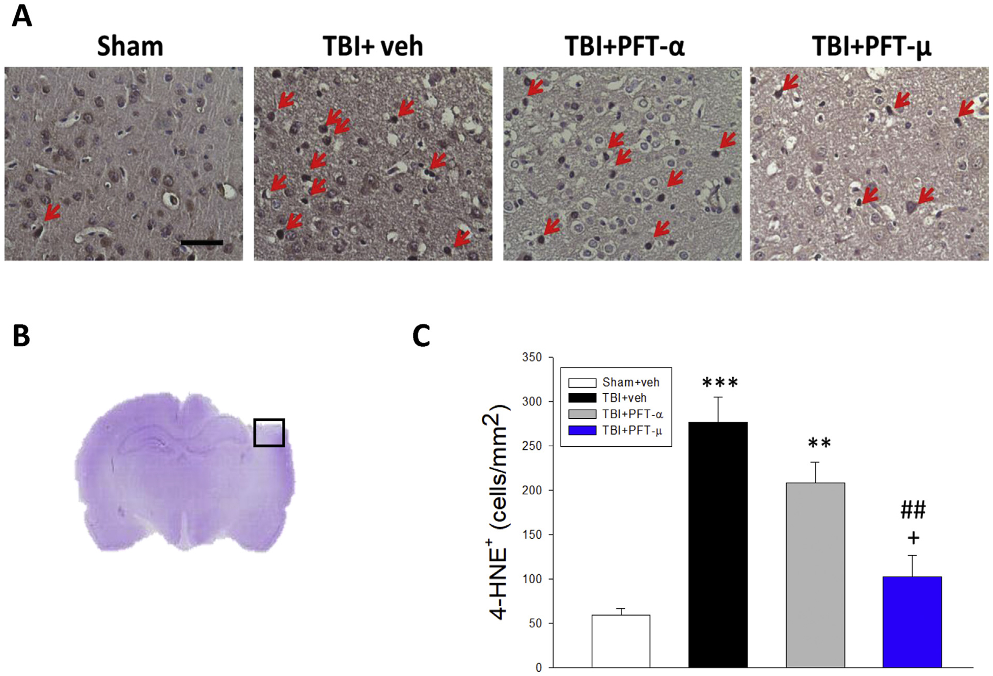
Fig. 6.

Post-injury administration of PFT-μ, but not PFT-α, at 5 h after TBI decreased 4-HNE positive cells in the cortical contusion region at 24 h. (A) Representative photomicrographs showing the results of immunohistochemical staining with anti-4HNE in 4 groups. Red arrows indicate 4-HNE-positive cells (brown colour). (B) A representative HE-stained coronal section showing the area as indicated by the black (ipsilateral cortex) square box to compare the fluorescent signals across the 4 groups of rats. (C) There was a significant decrease in the number of 4-HNE-positive cells in the TBI + PFT-μ group. The total number of 4-HNE-positive cells was expressed as the mean number of cells/mm2. Mean ± SEM. **P < .01, ***P < .001, compared with the sham group. ##P < .01, compared with the TBI + veh group. +P < .05, compared with the TBI + PFT-α group. (Scale bar = 50 μm; n = 5 per group).
