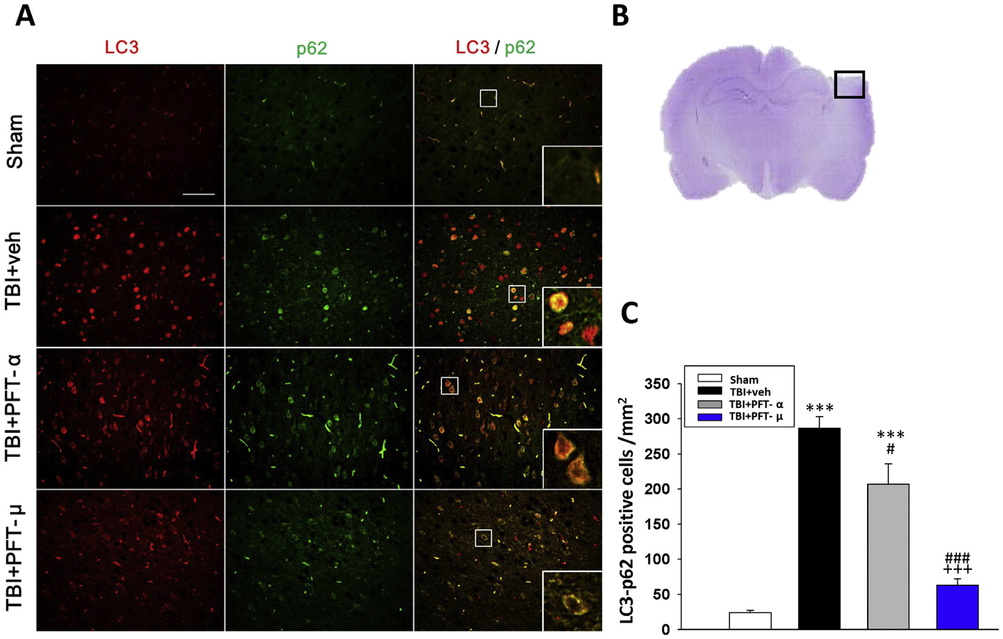
Fig. 7.

Post-injury administration of PFT-μ at 5 h after TBI decreases LC3/p62 positive cells in the cortical contusion region at 24 h. (A) Co-immunohistochemistry of LC3 and p62 in cortical brain tissue. LC3: green, p62: red, and yellow labelling indicating colocalization (Bar = 100 μm, 0.259 mm2). Inserts: Magnified colocalization of LC3 (red) and p62 (green) cells at a greater magnification. (B) A representative HE-stained coronal section showing the area as indicated by the black (ipsilateral cortex) square box to compare the fluorescent signals across the 4 groups of rats. (C) A significant decrease in LC3/p62 positive cells was evident in the TBI + PFT-μ group. The total number of LC3/p62-positive cells was expressed as the mean number per field of view in cortex. Data represent mean ± S.E.M. (n = 5 per group). **P < .01, ***P < .001 compared to the sham group; #P < .05, ### P < .001 compared to the TBI + veh group; +++ P < .001 compared to the TBI + PFT-α group.
