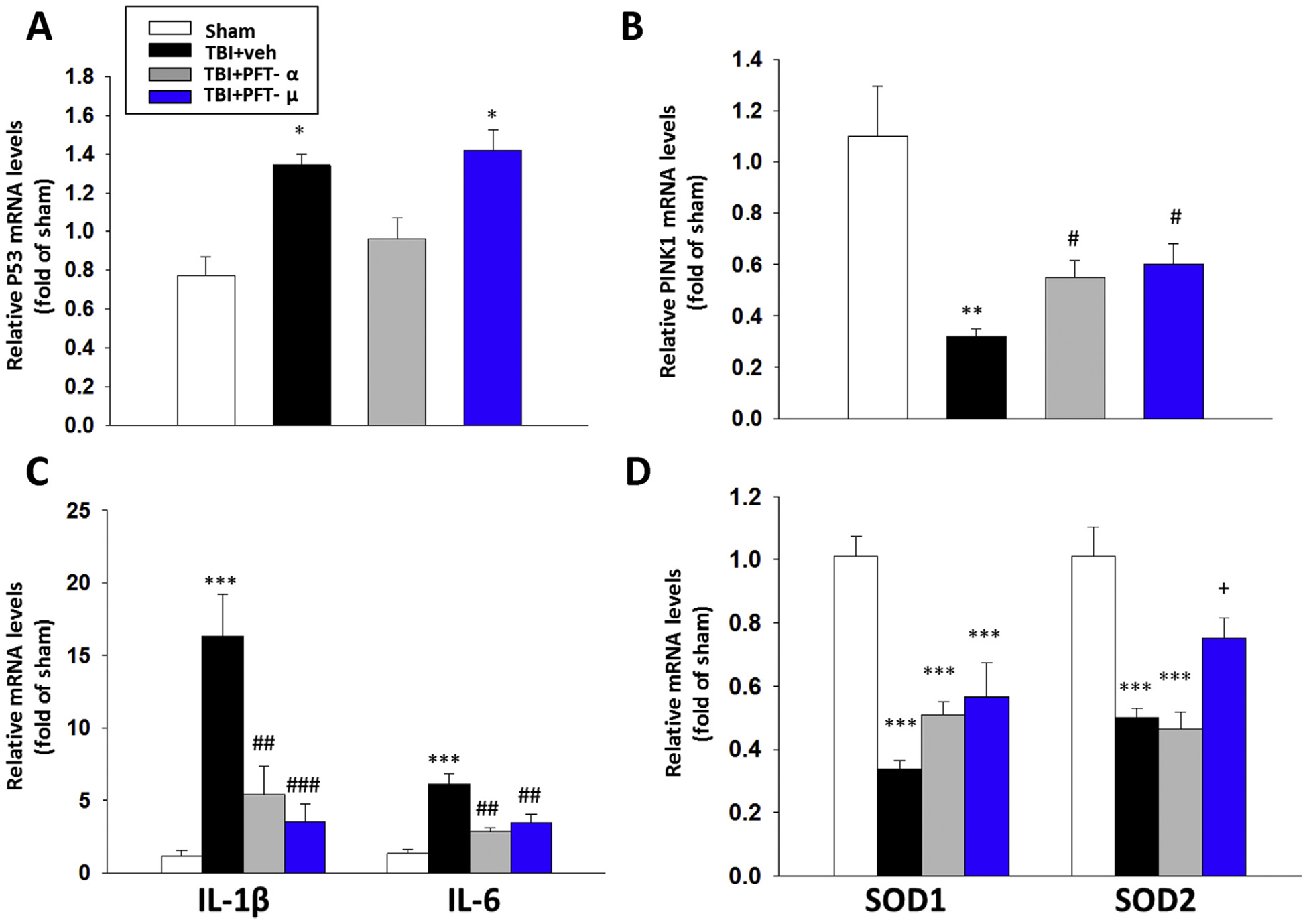
Fig. 8.

Treatment with PFT-μ at 5 h after TBI significantly mitigated TBI-induced reductions in PINK-1, and SOD2, and inhibited TBI-mediated rises in IL-1β, IL-6 mRNA expression in the cortical contusion region at 24 h. The mRNA levels of (A) p53, (B) PINK1, (C) IL-1β and IL-6, and (D) SOD1 and SOD2 across the four animal groups were evaluated by RT-PCR. PFT-μ significantly increased PINK1 and SOD2 mRNA expression, and decreased IL1β and IL-6 mRNA compared with TBI + vehicle-treated rats. Notably, PFT-μ did not change the level of p53 gene expression in accord with its transcriptional independent mechanism of action. Data are expressed as means ± SEM (n = 5 per group). * P < .05, **P < .01, ***P < .001 vs. sham; #P < .05, ##P < .01, ###P < .001 vs. TBI + veh group. +P < .05 vs. TBI + PFT-α group.
