Fig. 1.
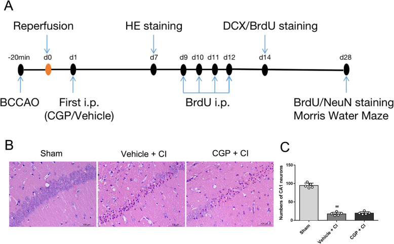
a The experimental protocol. The mouse CI model was induced by BCCAO for 20 min. Experimental mice were started to receive either CGP (10 mg/kg; i.p.) or an equal volume of vehicle (0.1 M PBS, i.p.) at day 1 after CI. HE staining was performed for morphological changes at days 7 after CI. BrdU (50 mg/kg; i.p.) was administrated daily for 4 consecutive days from 9 to 12 days after CI. And then mice were sacrificed for immunofluorescence staining (BrdU, DCX, NeuN) at 14 and 28 days after CI. Morris water maze (MWM) test was conducted to evaluate the spatial learning and memory abilities at days 28 after CI. b, c Effect of CGP on CI-induced histological changes in hippocampal CA1 neurons (n = 5 mice in each group). b Microphotographs of HE staining in hippocampal CA1 7 days after reperfusion (scale bar 100 μm). c Histogram showing the number of CA1 neurons in hippocampal (the data are expressed as the mean ± SD). ##P < 0.01 compared with the sham group (one-way ANOVA with Tukey post-test)

