Fig. 5.
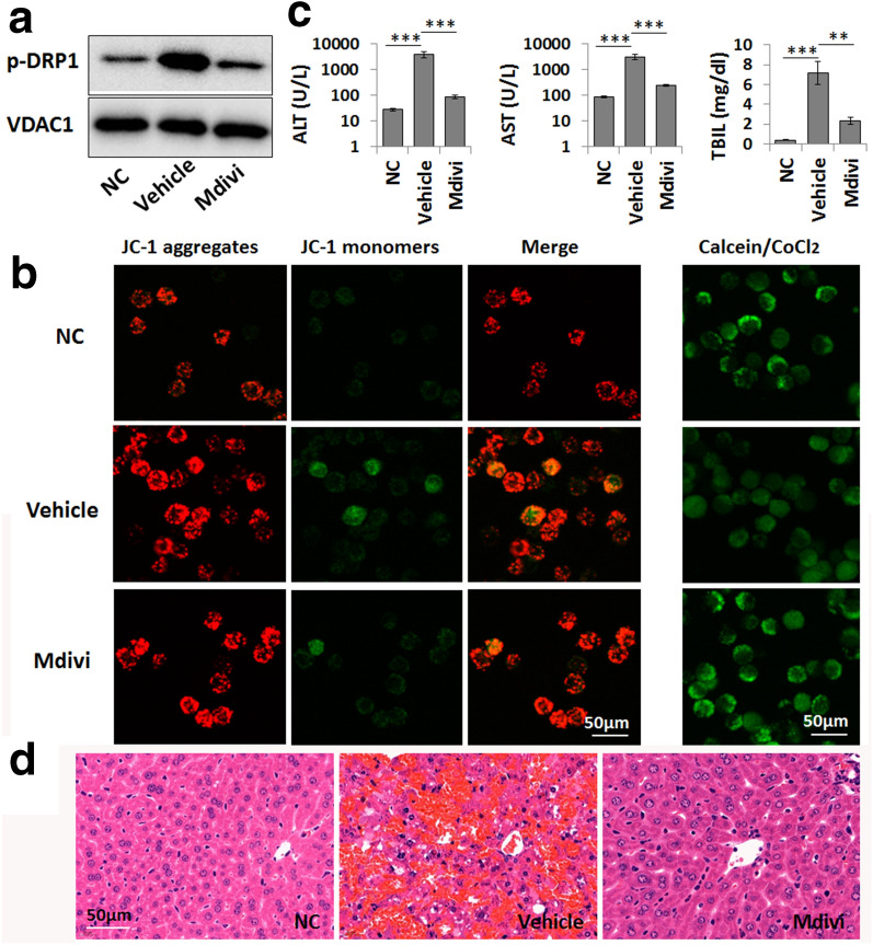
DRP1 mediates mitochondrial damage in macrophages in LPS/GalN-induced liver failure. a Western blot analysis of p-DRP1 levels in the mitochondrial fraction of normal cultured (NC) and LPS-primed macrophages. Mdivi (10 µM) was used to inhibit DRP1 activity. b The mitochondrial membrane potential (Δψm) and mitochondrial permeability transition pore (mPTP) opening in normal cultured (NC) and LPS-primed macrophages are shown by JC-1 (left) and calcein staining (right), respectively. c Serum levels of ALT, AST and TBIL in the normal control group (NC), vehicle-treated ALF group (Vehicle), and mdivi-treated group (Mdivi, 30 mg/kg). Data are presented as the mean ± SD (n = 6). **P < 0.01; ***P < 0.001. d Pathological changes in the liver tissue were shown by H&E staining

