Fig. 3.
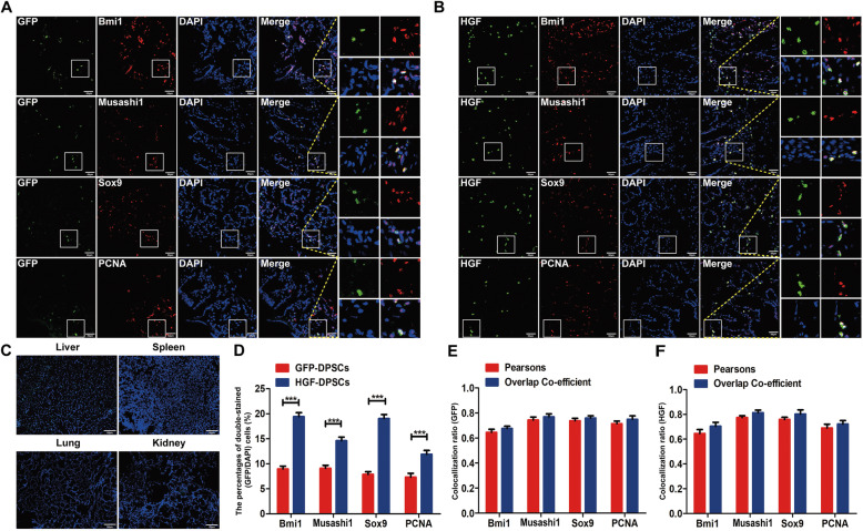
Transplanted DPSCs homed to injured colons and transdifferentiated into intestinal stem cell-like cells. a, b GFP-DPSCs and HGF-DPSCs expressing green fluorescence were colocalized with Bmi1, Musashi1, Sox9 and PCNA (n = 5). c Few positive cells were found in other organs of the rats (liver, spleen, kidney and lung tissues, n = 5). Scale bar = 50 μm in all panels. d Statistical comparison of the percentages of double-stained (GFP/DAPI) cells between the GFP-DPSCs and HGF-DPSCs groups. Data are shown as the means ± SD (n = 5; ***p < 0.001). e, f The comparison of Pearson’s correlation and the overlap coefficient of colon sections costained with Bmi1, Musashi1, Sox9 and PCNA between the GFP-DPSCs and HGF-DPSCs groups (n = 5; p > 0.05)

