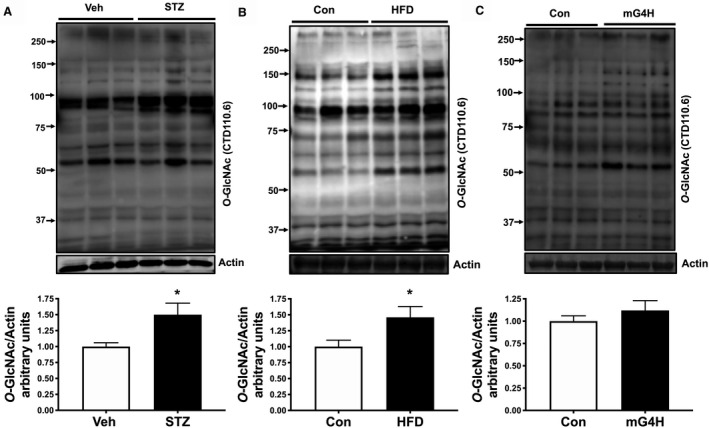
Figure 5. Protein posttranslational regulation via O‐GlcNAcylation is enhanced by STZ, HFD, and glucose.

Cardiac protein O‐GlcNAcylation was measured in ventricular lysates prepared from 4 weeks STZ‐induced diabetic mice, 12‐week HFD, or Con diet fed mice and mG4H mice after 2 weeks of transgene induction. Fifty micrograms of lysates was resolved and probed using CTD110.6 antibody. A, Upper panel: immunoblot illustrating O‐GlcNAc detection in ventricular tissue of STZ mice compared with vehicle‐treated mice. Lower panel: Densitometric analysis of immunoblots shown in upper panel. B, Upper panel: immunoblot illustrating O‐GlcNAc detection in ventricular tissue of HFD mice compared with vehicle‐treated mice. Lower panel: Densitometric analysis of immunoblots shown in upper panel. (C) Upper panel: immunoblot illustrating O‐GlcNAc detection in ventricular tissue of mG4H mice compared with vehicle‐treated mice. Lower panel: Densitometric analysis of immunoblots shown in upper panel. *P<0.05. Con indicates Control; HFD, high‐fat diet; MG4H, transgenic inducible cardiac‐restricted expression of glucose transporter 4; STZ, streptozotocin; Veh, Vehicle.
