Fig. 2.
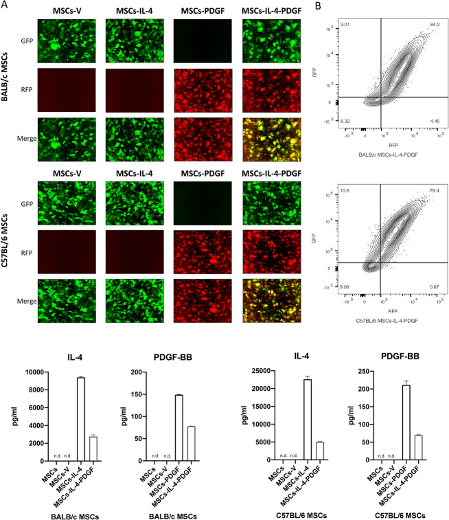
Characterization of infected MSCs by IL-4 and/or PDGF-BB lentiviral vectors and the expression level of IL-4 and PDGF-BB of these MSCs. a Representative fluorescence microscopy images of MSCs infected by lentiviral vectors. GM MSCs are GFP/RFP positive. b Characterization of MSCs-IL-4-PDGF groups, GFP, and RFP was analyzed by flow cytometry. c IL-4 and PDGF-BB secreting level detected by ELISA in the GM MSCs culture medium for 1-day culture. (n.d.: Cannot be detected by ELISA). “MSCs”, MSCs without infection; “MSCs-V”, MSCs infected with empty control lentivirus vector; “MSCs-IL-4”, MSCs infected with mIL-4 secreting lentivirus vector; “MSCs-PDGF”, MSCs infected with hPDGF-BB secreting lentivirus vector; “MSCs-IL-4-PDGF”, MSCs co-infected with both mIL-4 and hPDGF-BB lentivirus vectors, half of the dosage used in single infection respectively

