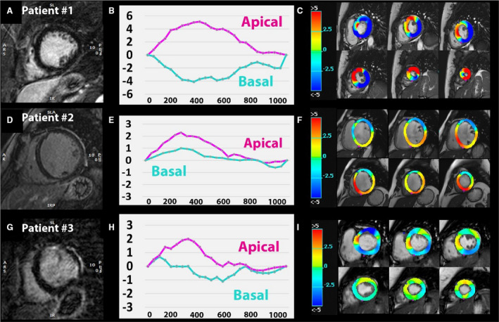
Figure 4. The location of LGE impacts LV mechanics.

Representative late gadolinium enhancement (LGE) images in 3 patients (first column A, D, and G), basal and apical circumferential rotation curves over time (second column B, E, and H), and circumferential rotation displayed as color‐coded parametric maps (third column C, F, and I). Patient 1 without LGE (A) with physiologic clockwise basal rotation (negative curve [B] and blue basal slices on circumferential displacement map [C]) and counterclockwise apical rotation (positive curve on [B] and red apical slices on [C]). In patient 2, midmyocardial septal fibrosis (D) and reversed basal rotation (positive curve on E) were detected with dominant counterclockwise motion on the color‐map (F). Patient 3 has free‐wall LGE (G) and shows impaired basal rotation (partly positive basal curve on [H] and light blue/green colors on [I]).
