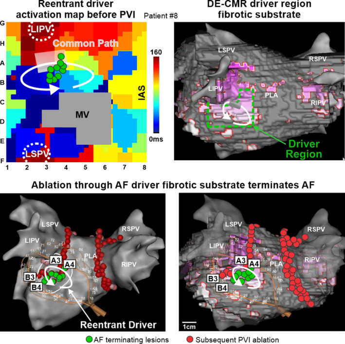
Figure 8. Targeted ablation of patient‐specific fibrotic reentrant driver substrate terminates AF.

Top Left, basket‐catheter activation map of a reentrant driver in the left atria (LA) of Patient #8. Top Right, delayed‐enhancement cardiac magnetic resonance (DE‐CMR) 3D reconstructions integrated with electroanatomic maps show the relationship between enhanced voxels (purple) and the analyzed driver region (dashed green box). Bottom, 3D electroanatomic map showing location of reentrant atrial fibrillation (AF) driver, AF terminating ablation lesion placement (green dots) and basket‐catheter position (left) and with integrated DE‐CMR reconstruction (right). Red dots denote subsequent pulmonary vein isolation. IAS indicates interatrial septum; LIPV/LSPV/RIPV/RSPV, left/right inferior/superior pulmonary veins; MV, mitral valve; PLA, posterior left atrium; and PVI, pulmonary vein isolation.
