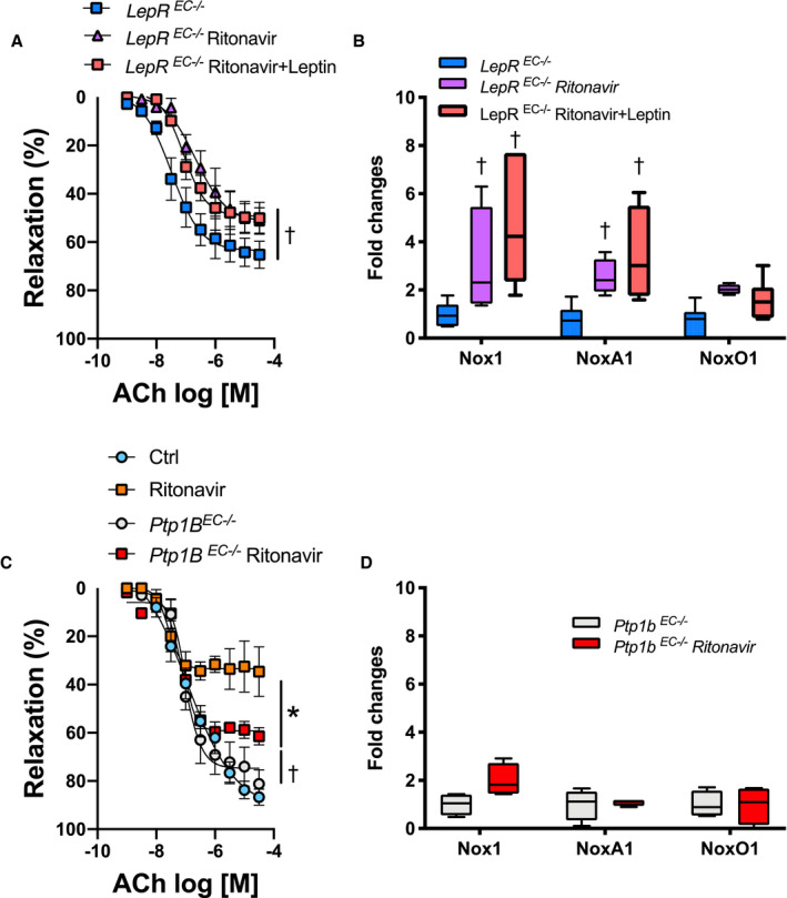
Figure 5. Leptin‐mediated vascular protection involves endothelial‐leptin signaling.

Concentration response curves to ACh in aortic rings (A and C) and real‐time polymerase chain reaction quantification of aortic NADPH oxidases subunits (B and D) in wild type, endothelial LepR–deficient (LepR EC−/−, A and B) or endothelial Ptp1B–deficient mice (Ptp1B EC−/−, C and D) treated with vehicle (Ctrl) or ritonavir (5 mg/kg per day for 4 weeks, ip) in the presence or absence of leptin treatment (0.3 mg/kg per day for 1 week, via osmotic mini‐pump). Vascular reactivity data are presented as mean±SEM. Gene expression data are presented as Min. to Max. N=3 to 8; *P<0.05 vs Ctrl; † P<0.05 vs LepR EC−/− or Ptp1b EC−/−. ACh indicates acetylcholine; Ctrl, control; LepR, leptin receptor; Nox1, NADPH oxidase 1; and Ptp1B, protein tyrosine phosphatase 1B.
