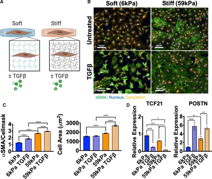
Figure 1. Cardiac fibroblast (CF) activation is modulated by culturing on different stiffness hydrogels and by adding TGFβ1 (transforming growth factor β 1) .

A, Schematic representation of experimental design to probe changes in myofibroblast activation in CFs. B, Representative images of immunofluorescence staining of CFs cultured for 5 days on hydrogels ±10 ng/mL TGFβ1 (green, α‐smooth muscle actin [αSMA]; yellow, cellmask; blue, nuclei; bar=100 μm). C, Changes in αSMA/cellmask intensity and changes in cell area quantified (N=individual cells from 2 experiments, error bars show 95% CI, 1‐way ANOVA, ****P≤0.0001). D, Quantitative reverse transcription–polymerase chain reaction measurement of periostin (POSTN) and transcription factor 21 (TCF21) expression in CFs on hydrogels±TGFβ1 (N=3, error bars show mean and SEM, 1‐way ANOVA, *P≤0.05, **P≤0.01, ***P≤0.001, ****P≤0.0001).
