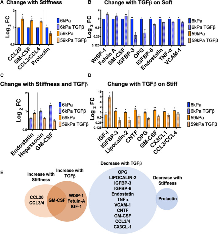
Figure 4. Cytokines that significantly increased or decreased in the conditioned media of cardiac fibroblasts (CFs) cultured on soft vs stiff hydrogels ±10 ng/mL TGFβ1 (transforming growth factor β 1).

A, Cytokines that change by culturing on different stiffnesses, *P≤0.05. B, Cytokines that change with TGFβ1 treatment on soft hydrogels, *P≤0.05, **P≤0.01. C, Cytokines that change between CFs grown on soft hydrogels with TGFβ1 vs CFs grown on stiff hydrogels with TGFβ1, *P≤0.05. D, Cytokines that change with TGFβ1 treatment on stiff hydrogels, *P≤0.05, **P≤0.01. E, Cytokines that significantly increased with myofibroblast activation (P≤0.05) are shown in orange and cytokines that decreased with activation are shown in blue. Overlap between treatment with stiffness and TGFβ1 is shown in the Venn diagrams. Cytokines that change with TGFβ1 treatment on either soft or stiff hydrogels are displayed in the Venn diagrams. All data are normalized to 6 kPa condition for each isolation. (N=3 isolations for 6 kPa, 59 kPa, 59 kPa TGFβ, N=2 for 6 kPa TGFβ, error bars show mean and SEM, 2‐tailed unpaired T test). FC indicates fold change; GM‐CSF, granulocyte‐macrophage colony‐stimulating factor; IGFBP, insulin growth factor binding protein; OPG, osteoprotegerin; TNF‐α, tumor necrosis factor‐α; VCAM‐1, vascular cell adhesion molecule 1; WISP‐1, Wnt1 inducible signaling pathway protein 1; CCL, Chemokine (C‐C motif) ligand; and CNTF, Ciliary neurotrophic factor.
