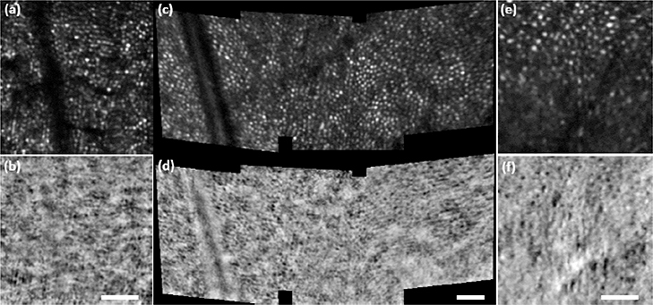
Fig. 5. Supine handheld imaging:
M-HAOSLO images acquired in handheld operation mode on supine dilated adult volunteers. (a) Averaged confocal image using 4 frames from S1. (b) Co-registered and averaged SD image from S1. (c) Four mosaiced, averaged confocal images using 6–8 frames each from S4. (d) Four mosaiced, co-registered and averaged SD images from S4. (e) Averaged confocal image using 4 frames from S3. (f) Co-registered and averaged SD image from S3. Scale bars for each image pair, 0.25°.

