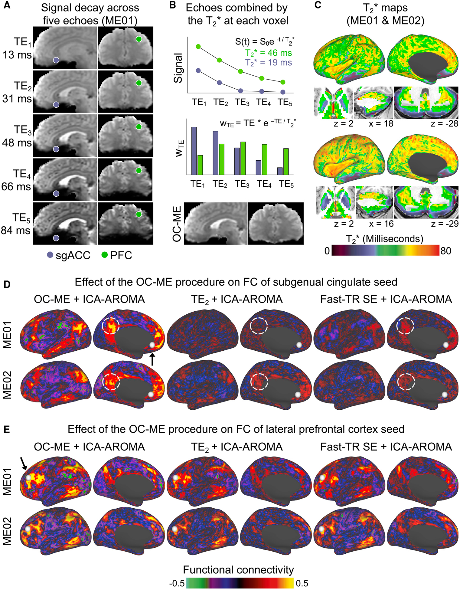
Figure 2. A Key Benefit of Multi-echo (ME) fMRI Is Improved BOLD Contrast and Reduced Signal Dropout after Echoes Are Combined.

(A) A ME fMRI sequence acquires multiple images at different echo times (TE) spanning dozens of milliseconds.
(B) Signals decay more rapidly in brain regions with a short T2* value, such as the subgenual anterior cingulate cortex (sgACC; purple). Echoes are combined such that those near the estimated T2* at each voxel are weighted most heavily, yielding an “optimally combined” ME (OC-ME) time series with improved BOLD contrast, less signal dropout, and dampened thermal noise.
(C) WTE represents the optimal weight for each echo. T2* values are calculated at each point in the brains of ME01 and ME02. Differences in the FC of seed regions with different T2* values help to convey the region-specific effect of the OC-ME procedure. FC maps were created using OC-ME and two different kinds of SE data (the second echo of the ME scan and a separate fast-TR SE sequence with a faster sampling rate) that were collected from both ME01 and ME02. PFC, prefrontal cortex; TE2, second echo of the ME scan.
