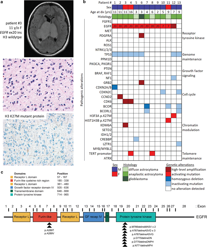
Fig. 1.
Genetic landscape of pediatric bithalamic gliomas. a, Axial T2-weighted fluid-attenuated inversion recovery (FLAIR) MR image from a 11-year-old girl (patient #3) showing an expansile mass involving the bilateral thalami (top). Histology from the stereotactic biopsy revealing a diffuse astrocytic neoplasm with pleomorphic nuclei and occasional mitotic figures (H&E stain, 400x magnification, middle). Negative immunohistochemical stain for histone H3 K27M mutant protein (400x magnification, bottom). b, Oncoprint table of the clinical features and likely pathogenic alterations identified in biopsies from thirteen children with bithalamic gliomas. Exon number where the EGFR mutations are located is annotated. c, Diagram of human EGFR protein with the location of the recurrent exon 20 small in-frame insertions within the intracellular tyrosine kinase domain and p.A289T/V missense mutations within the extracellular ligand-binding domain. UniProt P00533, RefSeq NM_005228.

