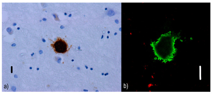
Figure 4.
Burnt-out plaques: (a) immunofluorescence visualization of a burnt-out Aβ plaque (the dense core remains) in an AD patient. Primary antibodies: anti-beta amyloid rabbit IgG. The original magnification was 400×. The scale bar indicates a length of 10 micrometers. (b) Imaging of the dense Aβ nucleus (green) lacking surrounding components using a confocal microscope. Primary antibodies: Anti-beta amyloid rabbit IgG and AT8 (murine anti-hyperphosphorylated protein tau). The secondary antibody was conjugated with either Alexa®488 (anti-rabbit IgG, green) or Alexa®568 (anti-mouse IgG, red). The scale bar indicates a length of 10 micrometers. This image comes from the amygdala of an 83-year-old female with a late variant of AD in the neocortical stage (Braak V, CERAD C). The changes associated with AD are at a “high” level (A3B3C3) according to the revised “ABC” classification of the NIA. Burnt-out and cored neuritic plaques were predominant in this area of the patient’s brain.

