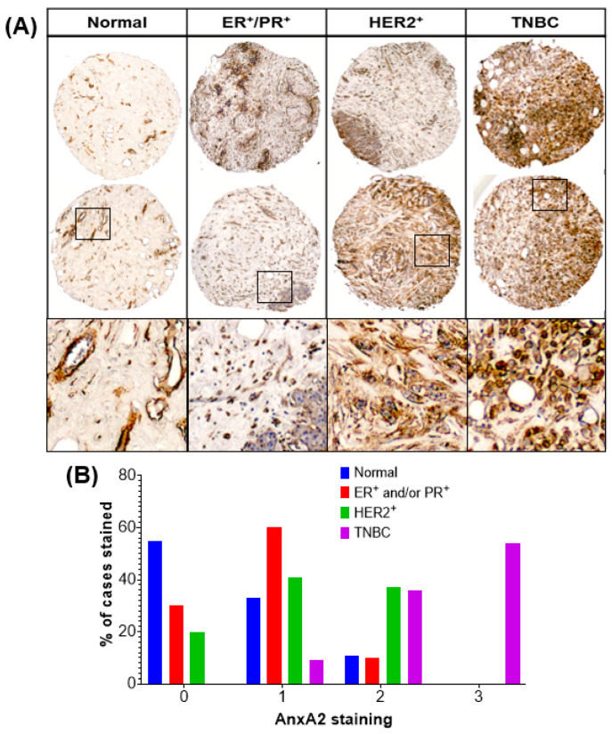
Figure 1.
Immunohistochemical analysis of AnxA2 expression in different subtypes of breast cancer tissue and normal breast tissue specimens. (A) Paraffin embedded tissue sections were stained with AnxA2 monoclonal antibody. Representative images of normal, ER+/PR+, HER2+, and TNBC tumor tissue specimens showing status of AnxA2 expression. AnxA2 was primarily localized to the plasma membrane of tumor cells in TNBC specimens. (B) Bar chart showing the AnxA2 staining patter in normal and different subtypes of breast cancer tissue specimens (Chi square test: χ² = 50.54, p < 0.0001).

