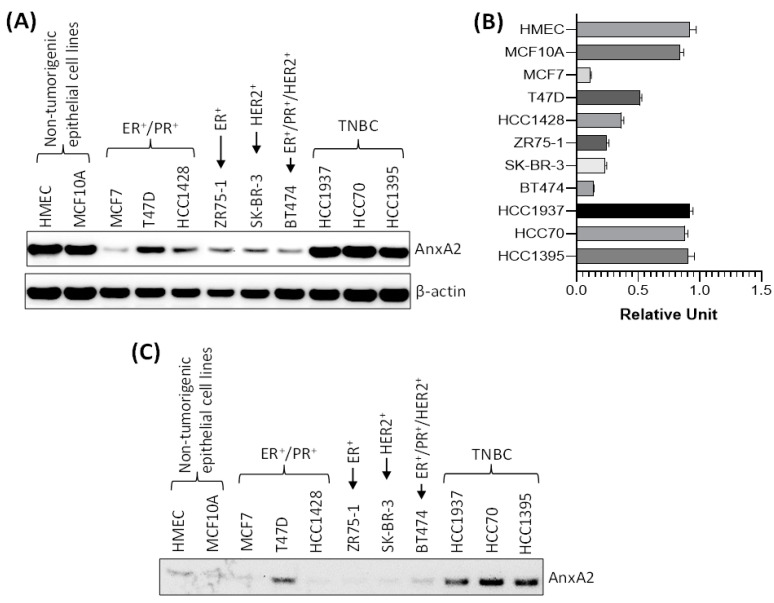
Figure 4.
Expression and secretion of AnxA2 in breast cancer cell lines. (A) AnxA2 protein expression was analyzed by immunoblotting in breast cancer cell lines and non-tumorigenic mammary epithelial cell lines. The membrane was stripped and reprobed with anti-β-actin antibody for loading control. (B) Bar chart showing the densitometric analysis (using ImageJ software) of AnxA2 bands of the immunoblot of panel A. Intensity of AnxA2 bands was normalized by β-actin loading control. Each bar represents the mean ± SE of three independent experiments. (C) Non-tumorigenic or breast cancer cells (6 × 10⁵) were plated in 100mm Petri dish for overnight and then switched to their respective serum free medium. After 24 h, AnxA2 was immunoprecipitated from the medium using anti-AnxA2 antibody and expression of AnxA2 secretion was analyzed by immunoblot analysis. Uncropped Western Blots of (A,C) are available in Figure S2.

