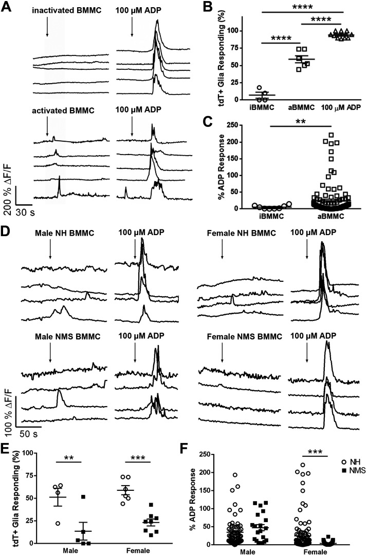
Fig. 2.
Mast cell supernatants evoke Ca2+ responses in myenteric glia. Data show Ca2+ imaging of myenteric glia in whole-mount preparations of colon isolated from Sox10CreERT2;GCaMP5g-tdT mice. A: representative traces showing glial Ca2+ responses as reported by GCaMP fluorescence (black traces) within a myenteric ganglion. Cells were challenged with supernatants obtained from cultures of bone marrow-derived mast cells (BMMC) at rest (inactivated, i) or following activation with DNP (activated, a). ADP (100 µM) served as a positive control. ΔF/F, change in fluorescence. B and C: quantification of the effects of BMMC supernatants on the percentage of glia [tdT-positive cells (tdT+)] responding to iBMMC, aBMMC, and ADP (B) and the percent of ADP response (C). D: representative traces showing glial Ca2+ responses as reported by GCaMP fluorescence (black traces) within a myenteric ganglion. Cells were challenged with activated supernatants obtained from cultures of BMMC from normally handled (NH) and neonatal maternal separation (NMS) male and female mice. ADP (100 µM) served as a positive control. ΔF/F, change in fluorescence. E and F: quantification of the effects of activated BMMC supernatants on the percentage of glia (tdT+ cells) responding to BMMC and ADP (E) and the percent of ADP response (F). Data are representative of recordings in n = 21–115 glial cells from five to eight ganglia from four mice. **P < 0.01, ***P < 0.001, ****P < 0.0001, Student’s t test and one-way and two-way ANOVA.

