Fig. 3.
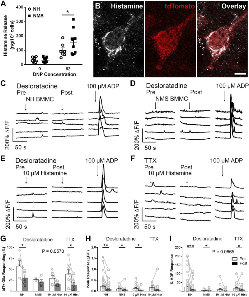
Histamine is a mediator of mast cell-glia communication. A: activated bone marrow-derived mast cells (BMMCs) [2×10⁶ cells/mL, stimulated with 0 and 62 ng/mL 2,4-dinitrophenol (DNP)] from neonatal maternal separation (NMS) mice release more histamine compared with activated BMMCs from normally handled (NH) mice. B: confocal images of the colon from Mcpt5Cre;GCaMP5g-tdT mice showing histamine immunoreactivity (grayscale) and tdTomato immunoreactivity (red) to mark mast cells. Scale bar (B, overlay) = 20 µm. C and D: representative traces of Ca2+ responses in myenteric glial cells evoked by exposure to activated BMMC supernatant from NH and NMS mice pre- and post-histamine type 1 receptor (H1R) antagonism with desloratadine (10 µM, left and right, respectively). E: representative traces of Ca2+ responses in myenteric glial cells evoked by exposure to histamine (10 µM) pre- and post-H1R antagonism with desloratadine (10 µM, left and right, respectively). F: representative traces of Ca2+ responses in myenteric glial cells evoked by exposure to histamine (10 µM) pre- and post-TTX (1 µM, left and right, respectively). G–I: quantification of the effects of H1R antagonism on the number of responding cells (G), peak glial Ca2+ responses evoked (H), and glial responses normalized to ADP responses (I). Data are representative of recordings in n = 17–63 glial cells from at least three ganglia from at least three mice and 7–16 BMMC cultures. *P < 0.05, **P < 0.01, ***P < 0.001, Student’s t test.

