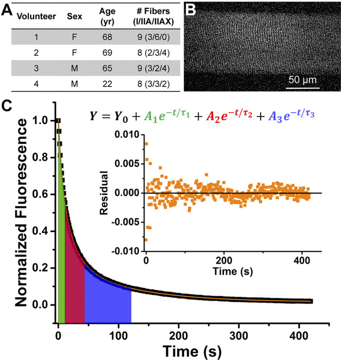
Fig. 1.

Measuring fluorescent nucleotide exchange activity of human skeletal muscle fibers. A: volunteer characteristics. B: representative fluorescence image of a single human skeletal muscle fiber loaded with mantATP before start of ATP exchange wash. C: representative normalized fluorescence trace (black) fitted with a 3-exponential decay function (orange). Color-shaded areas under the curve reflect the lifetime (τ) of each exponential decay component. Green: nonspecific washout; red: disordered-relaxed; blue: super-relaxed. Inset: residual plot from fitting function. mantATP, 2′/3′-O-(N-methylanthraniloyl) ATP.
