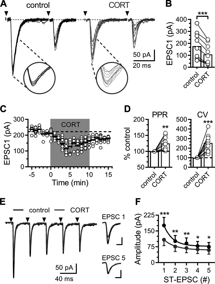
Fig. 1.
Corticosterone rapidly suppresses evoked glutamate release. A: representative solitary tract-excitatory post-synaptic currents (ST-EPSC) traces recorded in artificial cerebrospinal fluid (ACSF; control; black traces) and following bath application of corticosterone (CORT; 1 µM; gray traces). Each trace displays 10 trials overlaid (shock artifact has been removed) with arrows indicating the time of ST stimulation. Insets highlight the amplitude variability of EPSC1. B: plot of the average amplitude of the initial ST evoked EPSC (EPSC1). CORT decreases EPSC1 amplitude (n = 9 neurons/6 mice; P < 0.001, paired t test). C: plot of individual EPSC1 amplitudes (open circles) over time. Black lines represent average EPSC1 amplitude of 10 consecutive trials over 1 min, and gray shading indicates time of CORT exposure. D: plots of the average paired-pulse ratio (PPR; EPSC 2/1) and the coefficient of variance (CV) of EPSC1 normalized to baseline. CORT increases both the PPR (n = 9 neurons / 6 mice; P = 0.008, paired t test) and CV (P < 0.001, paired t test). E: representative trace showing ST-evoked EPSCs during a train of ST stimulations (five shocks at 20 Hz). Traces are an average of 10 consecutive trials. Scales for inset traces are 5 ms and 100 pA. F: curves for frequency dependent depression (FDD) of mean ST-EPSC amplitude across five ST shocks. CORT significantly decreased the FDD response (P < 0.001, two-way ANOVA), as well as the size of each ST-EPSC at each shock (P < 0.001; P = 0.001; P = 0.004; P = 0.026; P = 0.026; using Holm-Sidak multiple-comparison test). Values are expressed as means ± SE *P < 0.05, **P < 0.01, ***P < 0.001.

