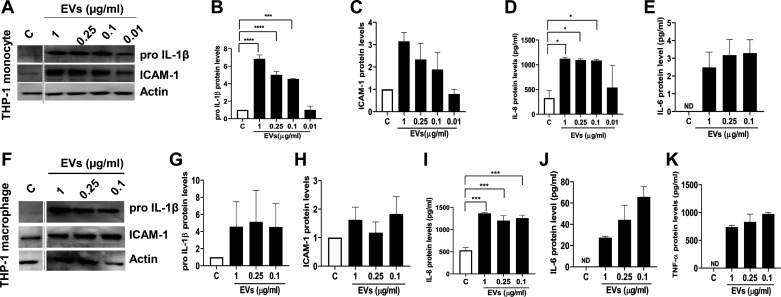
Fig. 6.
Effects of dust extracellular vesicles (EVs) on inflammatory mediator protein levels in THP-1 monocytes and THP-1 macrophages. THP-1 monocytes (A–E) and THP-1 macrophages (F–K) were untreated (C) or treated with medium containing different concentrations of EVs (μg/mL protein) for 5 h and the levels of proIL-1β, ICAM-1, and actin in cell lysates and the levels of IL-8, IL-6, and TNF-α in cell medium were determined. Noncontiguous lanes in Western blots are demarcated by broken lines. All data shown are means ± SE (n = 3). *P < 0.05, ***P < 0.001, and ****P < 0.0001 according to one-way ANOVA using Tukey’s multiple-comparison test. IL-6 and TNF-α levels in control medium were below detection [not detected (ND)].

