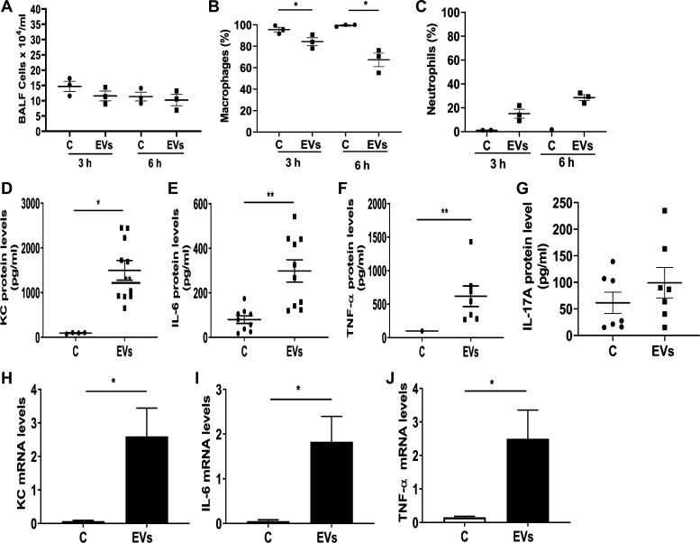
Fig. 9.
Effects of administration of dust extracellular vesicles (EVs) on inflammatory cell counts and inflammatory mediator protein levels in lungs of mice. Mice were administered PBS or dust EVs (1 μg protein) via intranasal instillation and bronchoalveolar lavage (BAL) fluid (BALF) was collected at 3 and 6 h after instillation and total RNAs from lung tissues were isolated. A–C: BAL fluid total cell counts were determined using a hemocytometer and macrophage and neutrophil counts were determined by Diff Quick staining. Percentages of macrophages and neutrophils in total BALF cells are shown. Data are means ± SE (n = 3). D–G: KC, IL-6, TNF-α and IL-17A levels in BALF samples collected at 3 h were determined by ELISA. Data shown are means ± SE (n = 7–10). H–J: KC, IL-6, and TNF-α mRNA levels in lungs collected at 3 h were determined by qRT-PCR. All data are shown as means ± SE (n = 6). *P < 0.05 and **P < 0.01 according to one-way ANOVA using Tukey’s multiple-comparison test.

