Fig. 11.
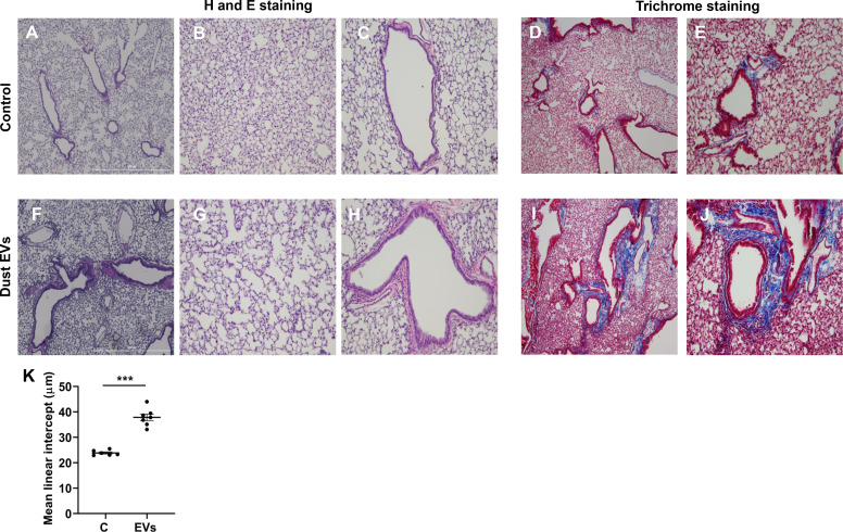
Effects of repeated administration of dust extracellular vesicles (EVs) on lung histology and collagen staining. Mice were administered 50 μL PBS or 50 μL dust EVs (1 μg protein) once daily (Monday–Friday) for ∼2 wk and lung sections were subjected to hematoxylin and eosin (H and E) staining. Collagen was visualized by trichrome staining. Representative images of H&E- and trichrome-stained lung sections from control and treated mice are shown at ×40 and ×100 magnification. A–C: H&E-stained sections from control mice. D and E: trichrome-stained sections from control mice. F–H: H&E-stained sections from dust EV-treated mice. I and J: trichrome-stained sections from dust EV-treated mice (n = 6 for control and n = 7 for EV-treated mice). K: images of lung sections from control and dust EV-treated mice were analyzed using ImageJ software to determine the mean linear intercept (Lm). Data shown are means ± SE (n = 6–7). ***P < 0.001 using t test.

