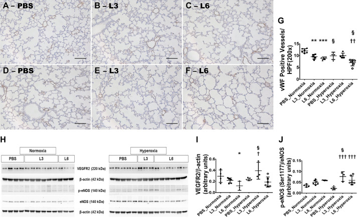
Fig. 4.
Lung vascularization deficits in 2-wk-old C57BL/6J wild-type mice exposed to LPS and hyperoxia during the saccular phase of lung development. C57BL/6J wild-type mice were exposed to either 21% O2 (normoxia) or 70% O2 (hyperoxia) during postnatal days (PNDs) 1–5 and injected intraperitoneally with 3 (L3) or 6 (L6) mg/kg of LPS or the vehicle (PBS) on PNDs 3–5. The mice were euthanized on PND 14 for lung morphometry and protein expression studies. A–F: Representative von Willebrand factor (vWF)-immunostained lung sections from mice treated with PBS (A and D), L3 (B and E), or L6 (C and F) and exposed to normoxia (A–C) or hyperoxia (D–F). Scale bar = 100 µm. Pulmonary vascularization was quantified by counting the number of vWF-stained lung blood vessels (G). VEGFR2, p-eNOS, eNOS, and β-actin protein levels were determined by immunoblotting (H). (I and J) VEGFR2 band intensities were quantification and normalized against β-actin (I) and p-eNOS against eNOS (J). Values represent the mean ± SD (n = 3–7 mice/group). Significant differences between PBS- and LPS-treated mice are indicated by *P < 0.05, **P < 0.01, and ***P < 0.001 under normoxic conditions and by †P < 0.05, ††P < 0.01, and †††P < 0.001 under hyperoxic conditions. Significant differences between treatment-matched mice under normoxic and hyperoxic conditions are indicated by §P < 0.05.

