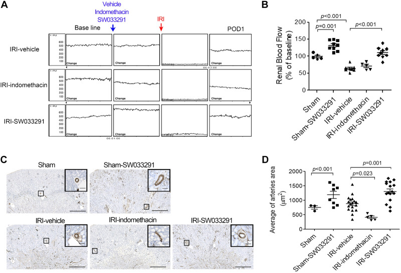
Fig. 4.
15-Hydoxyprostaglandin dehydrogenase inhibition induces renal vasodilation in the outer medulla of mice with ischemic acute kidney injury. To quantify vasodilation, the inner arteriolar area in the outer medulla was identified by α-smooth muscle actin staining. Assessments were performed at postoperative day 1 (POD1), 24 h after renal ischemia-reperfusion injury (IRI). A: representative images of the change in renal blood flow, as assessed by renal Doppler flux, with administration of vehicle, indomethacin, and SW033291. B: statistical analysis of renal blood flow in sham animals at time 0, in sham animals administered SW033291 (sham-SW033291) at 1 h postadministration of drug, and in cohorts subject to IRI and administered vehicle, indomethacin, and SW033291, which were then assayed at 24 h post-IRI. C: representative images of an arteriole in the outer zone of the renal medulla. Zoomed images are enlargements of the outlined areas. Scale bars in the enlarged images = 50 μm; scale bars in insets = 500 μm. D: statistical analysis of the inner arteriolar area of the outer medulla. Data are presented as means ± SE. Analysis was performed using Student’s t test.

